Back Muscles Anatomy Reference / Abdominal Muscle Description Functions Facts Britannica / While it is a strong muscle for adduction, internal rotation and extension of the humerus, we can do without it.
Back Muscles Anatomy Reference / Abdominal Muscle Description Functions Facts Britannica / While it is a strong muscle for adduction, internal rotation and extension of the humerus, we can do without it.. It is a sturdy, flat, triangular bone. Relevance of intrinsic muscles of the back for a fit pro. On this page, you'll learn about each of these muscles, their locations and functional anatomy. Superficial, intermediate, deep and deepest layers. Upper border of ribs ii to v.
They run along your spine, all the way from your sacrum up to your cervical spine. See more ideas about muscle anatomy, anatomy and physiology, massage therapy. The spine extends from the skull to the coccyxand includes the cervical, thoracic, lumbar, and sacral regions. The muscles of the back can be arranged into 3 categories based on their location: The deep back muscles are posterior to the erector spinae.
1 from The human back, also called the dorsum, is the large posterior area of the human body, rising from the top of the buttocks to the back of the neck. Muscles of the thoracic cage the muscles of the thoracic cage are the pectoralis major, pectoralis minor, serratus anterior, subclavius, intercostal (external, internal and innermost ), subcostal and transversus thoracis muscles. It is a sturdy, flat, triangular bone. Muscles of the back can be divided into superficial, intermediate, and deep group.since the all the back muscles originate in embryo (fetus) form by locations other than the back, muscles in the. These muscles stabilize the vertebral column and also have a role in proprioception and balance. The former two groups, superficial and intermediate, are referred to as the extrinsic back muscles. Lower back and superficial muscles. While it is a strong muscle for adduction, internal rotation and extension of the humerus, we can do without it.
Broadly considered, human muscle—like the muscles of all vertebrates—is often divided into striated muscle, smooth muscle, and cardiac muscle.
See more ideas about muscle anatomy, anatomy and physiology, massage therapy. However, the spinal erectors travel the length of the entire spine. This muscle is located on the upper portion of the back anatomy underneath the trapezius. To reduce complexity, muscle units have been incorporated in an abridged manner, reducing their actions more or less to a single force equivalent. Dimitrios mytilinaios md, phd last reviewed: Broadly considered, human muscle—like the muscles of all vertebrates—is often divided into striated muscle, smooth muscle, and cardiac muscle. Relevance of intrinsic muscles of the back for a fit pro. Muscle anatomy reference charts author: The tendon for latissimus dorsi can be harvested for tendon repairs or tendon transfers. The primary muscles for this are the multifidus and erector spinae. The deep back muscles, also called intrinsic or true back muscles, consist of four layers of muscles: The deep muscles of the back fit into or affix parts of themselves to the grooves in the spinous processes or the protrusion of the bone than can be felt through the skin. Make your life and learning a lot easier by using kenhub's muscle anatomy reference charts during your studies.
A basic understanding of the anatomy of your lower back can help you identify and differentiate a problem that commonly affects this region, such as localized muscle pain or sciatica. Latissimus dorsi is a large muscle that, when atrophied, can cause significance asymmetry in the back. Molly smith dipcnm, mbant • reviewer: Über 7 millionen englischsprachige bücher. The scapula provides attachment to several groups of muscles.
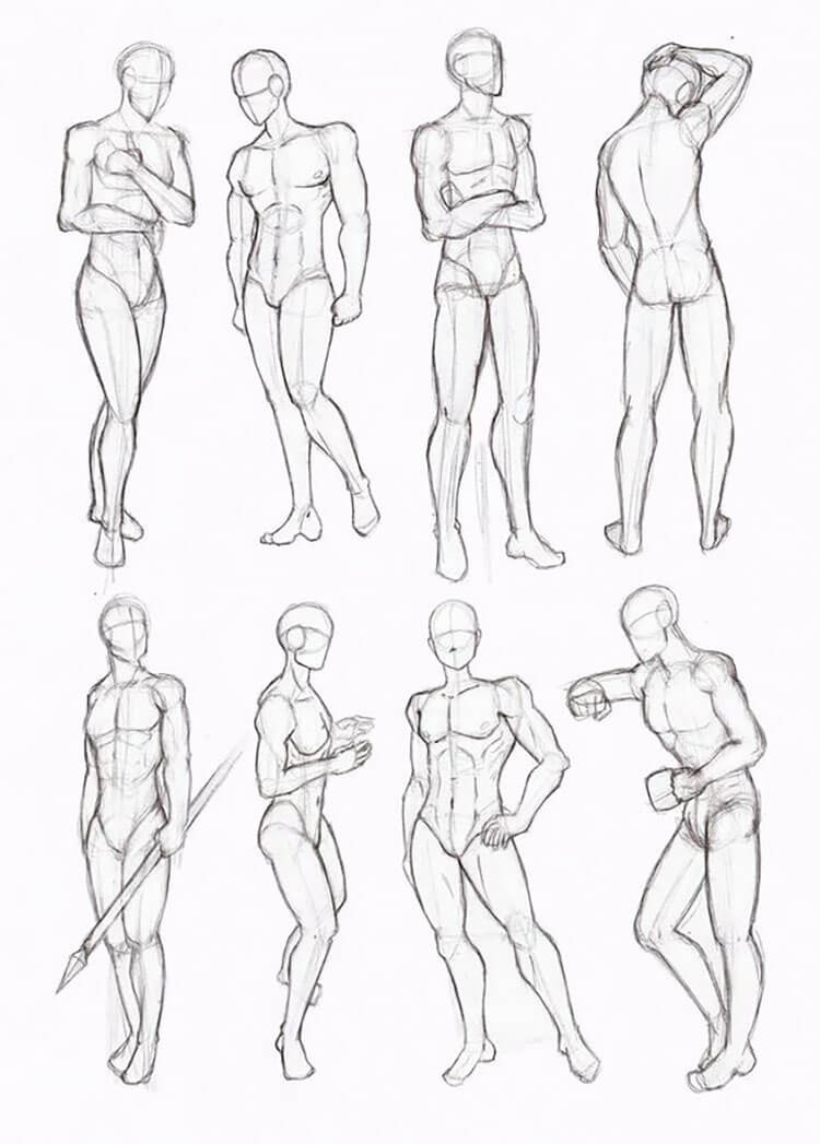
Learning The Muscles For Figure Drawing Love Life Drawing from www.lovelifedrawing.com Physicians, physiotherapists, athletes, students of sport, occupational therapists and Muscles of the thoracic cage the muscles of the thoracic cage are the pectoralis major, pectoralis minor, serratus anterior, subclavius, intercostal (external, internal and innermost ), subcostal and transversus thoracis muscles. These are the muscles that extend your spine or keeps it extended against loads, such as when you are doing a deadlift or a back extension. Learn how to draw the upper back muscles. The rhomboids are a collective group of muscles formed by the rhomboid major and minor. The spine extends from the skull to the coccyxand includes the cervical, thoracic, lumbar, and sacral regions. It is the surface of the body opposite from the chest and the abdomen.the vertebral column runs the length of the back and creates a central area of recession. Take free questions on this article.
Before we begin the tutorial, a.
The scapula or shoulder blade is the bone that connects the clavicle to the humerus. The muscles of the back are a group of strong, paired muscles that lie on the posterior aspect of the trunk. Muscles of the thoracic cage the muscles of the thoracic cage are the pectoralis major, pectoralis minor, serratus anterior, subclavius, intercostal (external, internal and innermost ), subcostal and transversus thoracis muscles. Take free questions on this article. Über 7 millionen englischsprachige bücher. Attached to the shoulder girdle. The primary muscles for this are the multifidus and erector spinae. The back muscles are anatomically layered into superficial (extrinsic) and deep (intrinsic) muscles. Muscle anatomy reference charts author: The extrinsic back muscles are located in the back, but act to produce movements of the shoulder and assist respiration. The spine extends from the skull to the coccyxand includes the cervical, thoracic, lumbar, and sacral regions. The scapula provides attachment to several groups of muscles. Upper border of ribs ii to v.
Make your life and learning a lot easier by using kenhub's muscle anatomy reference charts during your studies. Muscles of the back can be divided into superficial, intermediate, and deep group. The extrinsic back muscles are located in the back, but act to produce movements of the shoulder and assist respiration. The primary muscles for this are the multifidus and erector spinae. However, the spinal erectors travel the length of the entire spine.
21 Human Anatomy Drawing Ideas And Pose References Beautiful Dawn Designs from beautifuldawndesigns.net Muscle anatomy reference charts author: To reduce complexity, muscle units have been incorporated in an abridged manner, reducing their actions more or less to a single force equivalent. Largest muscle group in your body helps you burn more calories through the day! The scapula forms the posterior of the shoulder girdle. Before we begin the tutorial, a. They provide movements of the spine , stability to the trunk, as well as the coordination between the movements of the limbs and trunk. These muscles lie on each side of the vertebral column , deep to the thoracolumbar fascia . Attached to the vertebral column.
The intrinsic back muscles are found deeper to the extrinsic muscles, separated from them by the thoracolumbar fascia.
Anatomy_muscle_of_back 4/18 anatomy muscle of back train for maximum speed, power and accuracy. See more ideas about drawing reference, anatomy drawing, art reference poses. They are short muscles associated with the spinous and transverse processes of the vertebrae. The rhomboids are important in upper limb movement and stability of both the shoulder girdle and scapula. See more ideas about anatomy, anatomy reference, anatomy drawing. Über 7 millionen englischsprachige bücher. Ligaments hold the vertebrae in place, and tendons attach the muscles to the spinal column. The deep back muscles are posterior to the erector spinae. The rhomboids are a collective group of muscles formed by the rhomboid major and minor. The spine extends from the skull to the coccyxand includes the cervical, thoracic, lumbar, and sacral regions. On this page, you'll learn about each of these muscles, their locations and functional anatomy. The capitis and cervicis muscles, for example, generally originate on the thoracic spine. See more ideas about muscle anatomy, anatomy and physiology, massage therapy.
Back, followed by 144 people on pinterest back muscles reference. These muscles lie on each side of the vertebral column , deep to the thoracolumbar fascia .


0 Komentar